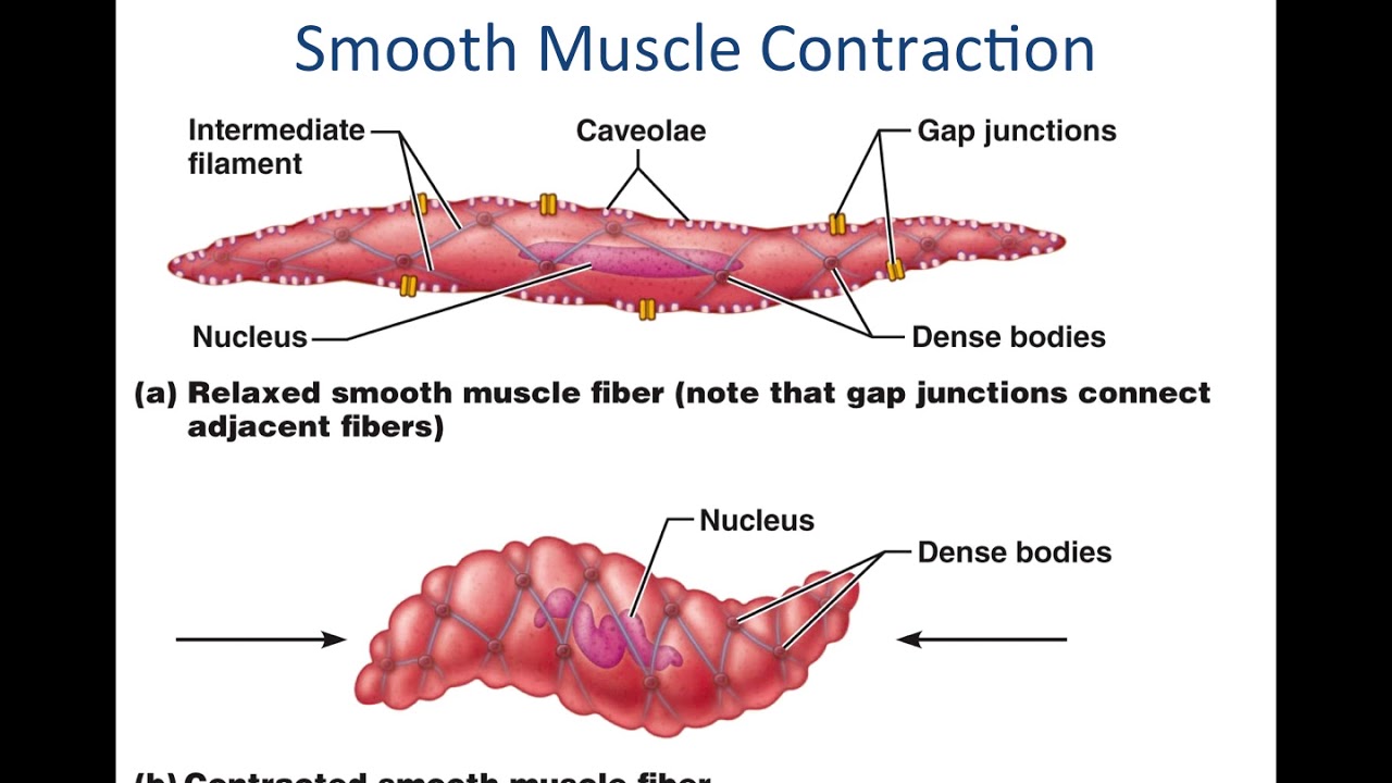
Smooth Muscle Diagram / This figure shows smooth muscle contraction. The left ... / There are 3 different types of muscle:. There are 3 different types of muscle: Smooth muscle tissue diagram labeled tissue photos and wallpaper upaaragon.co. 2 types of smooth muscle each fiber can contract independently and is usually innervated by a single nerve ending. Full color drawing pics 320x320 cardiac muscle and smooth muscle. • the new length however, retains its original _ seconds or minutes after it has been elongated or shortened (e.g.
Hair transplantation procedure diagram with steps. Keep reading to learn more about smooth muscle examples and how they function in the body. • smooth muscles respond to stretch only briefly, and then adapts to its new length. Smooth muscle histology and diagram (inlet). What is vascular smooth muscle?different types of muscle when we think of the word muscle, the image of bustling biceps or rippling abs may spring to mind.
Smooth Muscle Physiology - YouTube from i.ytimg.com Circuit diagram used for study 1141x1080 draw the diagram of smooth muscles or neuron muscle It is layered in a distinctive pattern of circular layers. This diagram depicts visceral smooth muscle and explains the details of visceral smooth muscle. Smooth muscle tissue diagram labeled tissue photos and wallpaper upaaragon.co. Smooth muscle tissue is also known as visceral muscle tissue. Smooth muscle is found in the walls of hollow organs like your intestines and stomach. Smooth muscle is a type of tissue found in the walls of hollow organs, such as the intestines, uterus and stomach. Smooth muscles are mainly divided into two subgroups:
It constitutes much of the musculature of.
Fibers insulated from each other by covering of collagen and glycoprotein fibrillae. What is vascular smooth muscle?different types of muscle when we think of the word muscle, the image of bustling biceps or rippling abs may spring to mind. Smooth muscle is found in the walls of hollow organs like your intestines and stomach. Smooth muscle lines the inside of blood vessels and organs, such as the stomach, and is also known as visceral muscle. Smooth muscle structure, embryonic origin, and histology. Flat vector design element for infographic poster. You can download and read online pdf file book smooth muscle diagram only if you are registered here.download and read online smooth muscle diagram pdf book file easily for everyone or every device. This is in contrast to skeletal and cardiac muscle, which have. Muscles in your bladder wall contract to expel urine from your body. They work automatically without you being aware of them. • the new length however, retains its original _ seconds or minutes after it has been elongated or shortened (e.g. Smooth muscle is found in the walls of hollow organs like your intestines and stomach. They are present iris of eye, in bronchi of lungs, alimentary canal drag the labels onto the diagram to label the steps of smooth muscle activation and deactivation.
Smooth muscle fibers ____x smaller than fibers in skeletal muscle. Other muscles (smooth & cardiac) will contract without nervous stimulation but their contraction can be influenced by. Smooth muscle is found in the walls of hollow organs like your intestines and stomach. Smooth muscle lines the inside of blood vessels and organs, such as the stomach, and is also known as visceral muscle. Key products for cardiovascular primary cell culture.
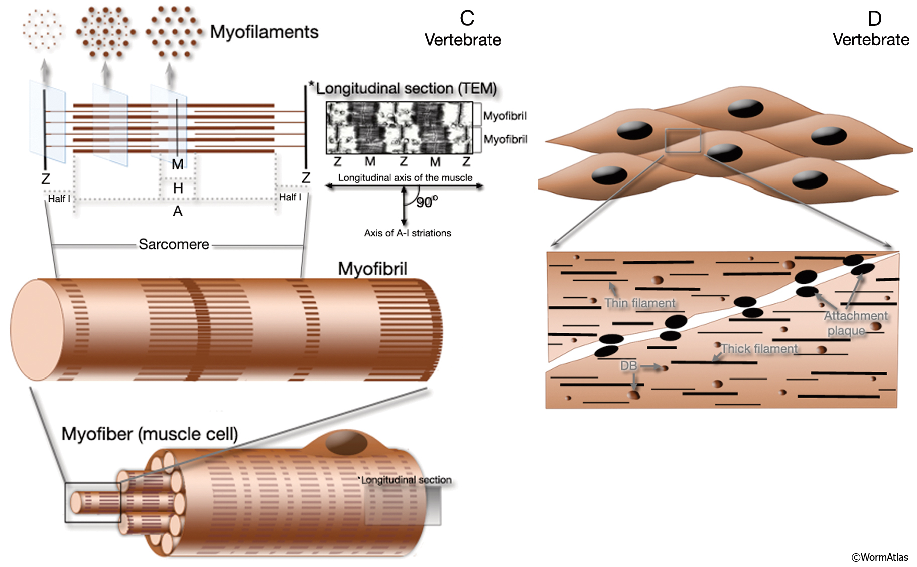
MusFIG 1C&D Legend from www.wormatlas.org Smooth muscle is also called involuntary muscle or unstriated muscle. Smooth muscle structure, embryonic origin, and histology. Diagram of artery with smooth muscle identification. Smooth muscle tissue is also known as visceral muscle tissue. Circuit diagram used for study 1141x1080 draw the diagram of smooth muscles or neuron muscle It is layered in a distinctive pattern of circular layers. The term smooth muscle refers to a muscle of the human body that is part of an involuntary muscle group. Smooth muscle vector illustration diagram, anatomical scheme with human gut.
• the new length however, retains its original _ seconds or minutes after it has been elongated or shortened (e.g.
This page describes smooth muscle development, descriptions of cardiac muscle and smooth muscle development can be found in other notes. Smooth muscle is also called involuntary muscle or unstriated muscle. Ciliary muscle of eye, iris, piloerector muscles. You can download and read online pdf file book smooth muscle diagram only if you are registered here.download and read online smooth muscle diagram pdf book file easily for everyone or every device. Keep reading to learn more about smooth muscle examples and how they function in the body. They work automatically without you being aware of them. Muscle diagram for chest and back. This diagram depicts visceral smooth muscle and explains the details of visceral smooth muscle. Smooth muscle lines the inside of blood vessels and organs, such as the stomach, and is also known as visceral muscle. Smooth muscle has a fusiform shape, which resembles a football or spindle. You will have some basic understanding of the appearance referring to the below smooth muscle diagram. Smooth muscle anatomy and physiology i. • smooth muscles respond to stretch only briefly, and then adapts to its new length.
Hair transplantation procedure diagram with steps. You can also find smooth muscle in the walls of passageways, including arteries and veins of de cardiovascular system. As in cardiac muscle cells, the configuration of the nuclear. Smooth muscles are mainly divided into two subgroups: Smooth muscle fibers do not have their myofibrils arranged in strict patterns as in striated muscle, thus no distinct striations are observed in smooth muscle cells under the microscopical examination.
Human Uterine Smooth Muscle Cells (HUtSMC) | PromoCell from www.promocell.com Smooth muscle fibers ____x smaller than fibers in skeletal muscle. They are present iris of eye, in bronchi of lungs, alimentary canal drag the labels onto the diagram to label the steps of smooth muscle activation and deactivation. Diagram of artery with smooth muscle identification. Smooth muscle histology and diagram (inlet). Human circulatory system vector illustration diagram, blood vessels scheme. It constitutes much of the musculature of. In this video i have shown the simplest way of drawing muscle drawing. This is different from cardiac muscle tissue, which develops into an as you look at this diagram of a smooth muscle fiber, you'll notice the single nucleus in the center.
Smooth muscle is under involuntary control and is innervated by the autonomic nervous system.
Muscle paintings search result at paintingvalley.com. Smooth muscle histology and diagram (inlet). This is in contrast to skeletal and cardiac muscle, which have. Smooth muscle, muscle that shows no cross stripes under microscopic magnification. Smooth muscles are mainly divided into two subgroups: The muscular walls of your intestines contract to push. Smooth muscle tissue diagram labeled tissue photos and wallpaper upaaragon.co. 2 types of smooth muscle each fiber can contract independently and is usually innervated by a single nerve ending. It is the pen diagram of skeletal, smooth and cardiac muscle for class 10, 11 and 12. This is different from cardiac muscle tissue, which develops into an as you look at this diagram of a smooth muscle fiber, you'll notice the single nucleus in the center. Vascular smooth muscle refers to the particular type of smooth muscle found within, and composing the majority of the wall of blood vessels. Smooth muscle is a type of tissue found in the walls of hollow organs, such as the intestines, uterus and stomach. • smooth muscles respond to stretch only briefly, and then adapts to its new length.


0 Komentar